
This is a 15 year old, quarter-horse, gelding with a 3 day history of acute onset right hind lameness.
At the time of the initial injury he was toe-touching lame on the right hind, but at the time of examination, he was 3.5/5 lame.
Examination revealed a large effusion (increased fluid within the joint) of the femoropatellar and medial femorotibial joints (two joints of the stifle). An upper limb flexion test on the right hind produced severe pain.
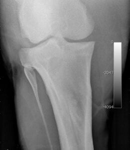
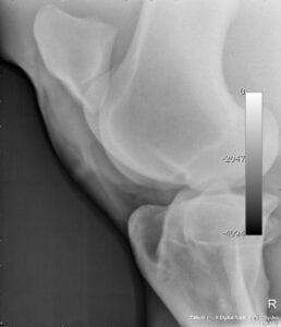
X-Rays
Digital X-rays were taken of the right stifle, but showed only a small osteophyte (bone spur) on the femur.
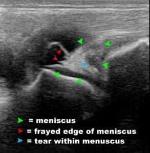
Ultrasound
The next step was to image the soft tissues of the stifle, including the meniscus, collateral ligaments, and joint cartilage via ultrasound. See image below for findings.
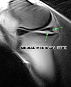
Stifle MRI
Recommended Treatment
Based on the imaging, we recommended arthroscopic surgery followed by regenerative therapy and rehabilitation.