Day: June 5, 2020

Why an Equine MRI?

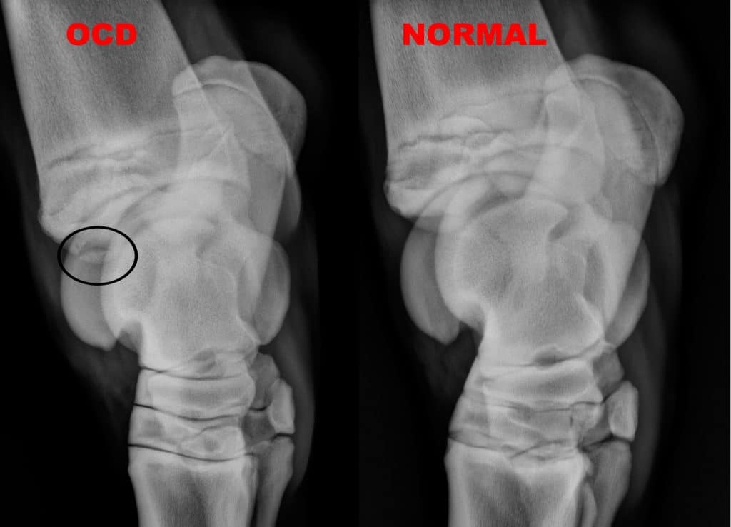
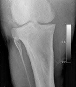
The number one reason for needing an MRI scan is equine lameness. There are many factors that can cause this painful condition, such as issues with the soft tissue structure and bone injuries. Diagnosing lameness requires a keen understanding of equine anatomy and physiology, conformation

Read More »
Oblique view of neck spinal cord compression.

Equine Wobblers

An equine wobbler is a horse with a damaged spinal cord. This can occur from malformation of the vertebral column, advanced arthritis in the vertebral joints or injury to the vertebrae. Any of these conditions can cause pressure on the spinal cord and cause the flow of

Read More »
Here's a short alt tag for the image: `X-ray showing bone spurs and joint damage`

Sand Enterotherapy

Sand Enteropathy is the accumulation of sand or gravel in the large colon, that subsequently causes inflammation of the colon wall. There is no doubt that most horses ingest some sand or gravel in their daily ‘graze’ around their stall or turn-out. I some areas

Read More »
MRI of suspensory ligament origin tear.

Suspensory Ligament

This is a 13 year old warmblood dressage horse that had had a previous right hind limb suspensory ligament injury 1.5 yrs. earlier. He returned to work, but developed recurrent right hind limb lameness when he advanced to a more demanding work level. He blocked

Read More »
Equine stifle MRI, sagittal view.

Navicular Syndrome

Pain that originates from the back of the foot or caudal heel pain. Recently, the definition of ‘heel soreness’ or ‘navicular syndrome’ in horses has been thrown on its head. With new developments in diagnostic tools for horses, it has become obvious that there are

Read More »
OCD and normal equine joint x-rays.

Hock OCD (Osteochondritis Dessicans)

OCD of the Distal Intermediate Ridge of the Tibia What is OCD of the distal intermediate ridge of the tibia? The most common joint in which to diagnose osteochondritis dissecans (OCD) in the horse is the tarsus. The most common location for OCD is the

Read More »
Here's an alt tag for the image: `X-ray of a horse's leg`

Meniscus Tear MRI

This is a 15 year old, quarter-horse, gelding with a 3 day history of acute onset right hind lameness. At the time of the initial injury he was toe-touching lame on the right hind, but at the time of examination, he was 3.5/5 lame. Examination

Read More »