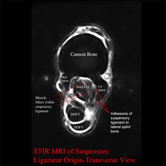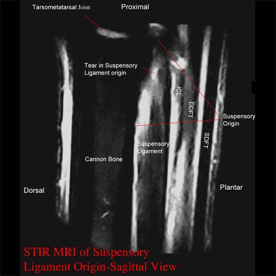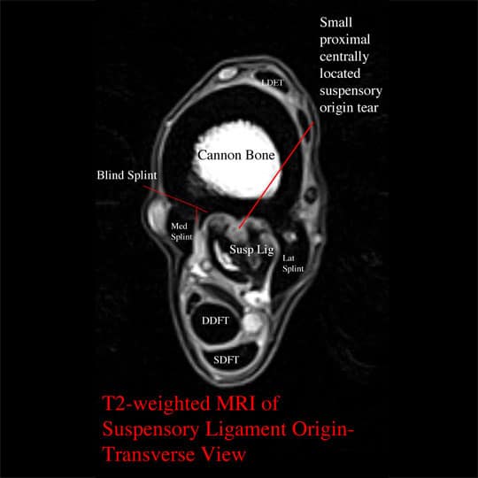
This is a 13 year old warmblood dressage horse that had had a previous right hind limb suspensory ligament injury 1.5 yrs. earlier. He returned to work, but developed recurrent right hind limb lameness when he advanced to a more demanding work level. He blocked sound with a suspensory origin block of the right hind limb. Digital Ultrasound, using a 13 MHz tendon transducer, showed no significant abnormalities of the suspensory ligament initially. MRI of the right hind limb suspensory ligament was performed: small centrally located, very proximal suspensory ligament origin tear (seen best on the STIR images), extensive adhesions of the suspensory ligament origin/body/branches to the medial and lateral splint bones and cannon bone, and multiple small “blind†splints were found impinging on the suspensory ligament. Post-MRI digital ultrasound could not identify most of the adhesions and “blind†splints, but with extensive searching found the small proximally located suspensory origin tear.
Treatment
This horse was treated with surgical takedown of adhesions of the suspensory ligament to the splint bones and cannon bone, and removal of multiple, small blind splints. Sepra-Film (hyaluronic acid paper) was placed between bone and suspensory ligament prior to closure of the incisions, to help prevent recurrent adhesions. The small, proximal suspensory ligament tear was injected with fat-derived stem cells. The horse returned to walking under saddle 6 weeks after surgery. At 3 months after surgery, the suspensory ligament tear appeared completely healed by digital ultrasound examination, and the horse was sound. The horse was allowed to return to work at that time and has remained sound since.