Equine Lameness Evaluation
At Cave Creek Equine® Sports Medicine & Surgery, our specialized board-certified veterinarians evaluate and treat lameness daily. For high-performance horses, subtle lameness can indicate underlying issues.
Every horse is unique. Identifying what triggers lameness is essential for diagnosing and treating the condition, particularly for equine athletes, where training and competition vary greatly depending on the sport.
Here’s what you can expect during a lameness evaluation at Cave Creek Equine® Sports Medicine & Surgery:
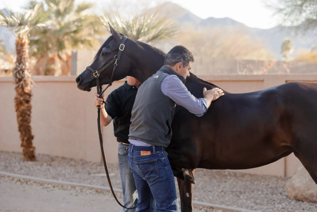
When a horse arrives, we first listen to the owner describe their observations and the conditions under which the horse has demonstrated lameness symptoms. We observe the horse at rest, at the walk, trot, and canter, where required. Depending on the situation, we will have the owner ride the horse so that we can evaluate the horse and rider as they function together.
We then perform a hands-on examination of the horse. During this lameness exam, we palpate the tendons, ligaments and joints, and perform various upper and lower limb flexion tests on all four legs. We trot the horse after each flexion exam to observe any changes in the horse’s gait.
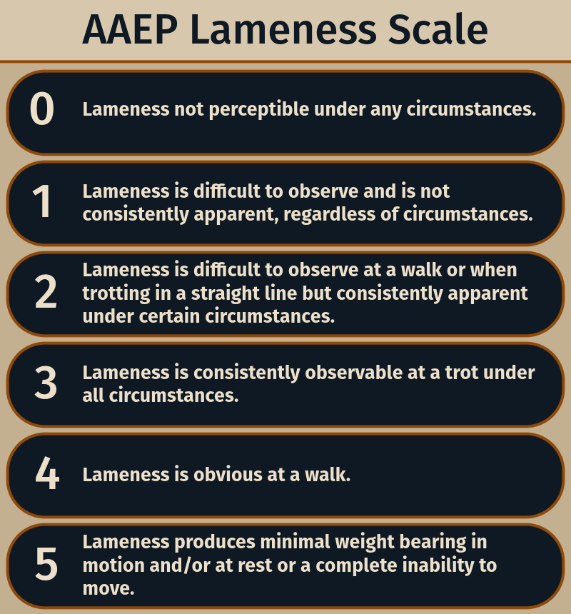
The American Association of Equine Practitioners (AAEP) developed this lameness grading system to aid both communication and record-keeping.
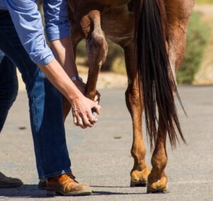
Once we’ve uncovered the approximate origin of lameness, we start at the hoof and inject an anesthetic nerve or joint block which numbs the foot. Then, we test the horse’s gait again. If there are no longer signs of lameness in the horse, we’ve now isolated the location of the lameness. If the nerve block does not improve the gait, we move up higher on the leg until we find the exact location of lameness.
The most common equine injuries are typically tears in tendons or ligaments, or joint injuries.
The location of the lameness influences the type of diagnostic tool we use. For instance, an Equine X-ray (digital radiography) is used for areas such as the joints and bones and shows us subtle lesions that might be causing the discomfort. An ultrasound (ultrasonography) can show us inflammation and tears in the soft tissues such as muscles, tendons, and ligaments.
The challenge we face is that neither the X-ray nor ultrasound can penetrate the hoof wall and therefore do not enable us to view the small complex structures in the foot. Using advanced imaging such as an Equine MRI, we typically find multiple injuries within the foot, which is understandable as the enormous forces acting on the foot dissipate into all of its tissues.
This limitation of X-ray or ultrasound also applies to very large joints such as the stifle joint, which are difficult to examine due to the large muscles surrounding them. Some soft tissues, such as the suspensory ligament, are also difficult to image because their architecture (partly made up of fat and muscle) is much more complex than that of tendons and other ligaments.
For example, in the images below, digital X-rays of the right stifle showed only a small bone spur on the femur. Based on the advanced imaging, we diagnosed the meniscal tear.
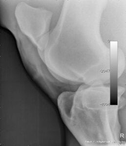
X-Ray
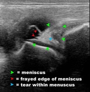
Ultrasound
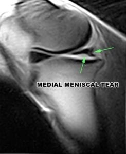
MRI
The MRI is the gold-standard for soft tissue evaluation. We are highly skilled and trained in MRI scanning and understand the value of longer scan times to produce high-quality, highly detailed images. That is why we purchased our Esaote Vet-MR scanner. A standing MRI provides substandard imaging quality due to short scan times and software that tries to correct for the motion of a standing horse. With our Esaote MRI scanner, because the horse is anesthetized, we’re able to ensure no movement can degrade image quality while ensuring the horse’s safety.
For injuries or issues in the bone, the nuclear scintigraphy or bone scan is an important imaging technique that allows us to examine active, and often subtle, bone injury or response under large muscle cover or along the spinal cord and pelvis. We commonly use bone scans to look for stress fractures, bone infraction, infections, and especially, arthritis of joints such as the facet joints between the cervical vertebrae in the neck of the horse.
Slow motion video analysis is another tool that is very helpful to assess subtle lameness issues in a horse. It is an invaluable tool to reliably assess and monitor the response to nerve and joint blocks as well as treatment and rehabilitation.
Barrel Racing
Dressage
Shoeing
Cutting
With this level of imaging and our expertise, we’re able to create a clear treatment plan for your horse’s lameness.
We are firm believers in regenerative therapies, where applicable, minimalizing invasive treatment. Dr. Vidal and Dr. Aristizabal are pioneers in equine stem cell research and administration and there are thousands of success stories regarding the anti-inflammatory effects and accelerated healing provided by stem cell therapy.
For example, PRP therapy harnesses the horse’s natural healing process by delivering a concentrated dose of platelets to an injury site. These platelets, the smallest blood cells, play a crucial role in clotting and tissue repair, acting as first responders to injury. IRAP is an anti-inflammatory protein that counteracts the destructive effects of inflammatory proteins using the horse’s own blood to stimulate white blood cells.
If required, our skilled equine surgeons will explain surgical options to you.
As you would expect, each of these steps is unique to your horse, their lameness symptoms, and your plans for the horse in terms of activity. We work with you every step of the way to ensure that your horse receives the best care possible.
We know you want the best for your horse so we’ve equipped our center with the latest diagnostic and imaging tools and have highly skilled, double-board certified equine vets focused on your horse’s well-being. We specialize in providing a thorough and systematic approach to equine lameness.
We treat your horse as if it’s our own. Cave Creek Equine® Sports Medicine & Surgery focuses on getting you an accurate diagnosis quickly, saving you time and money, and most important, saving your horse from pain.
Keep your equine athlete in peak condition. Schedule a consultation with our lameness specialists today.