WHY CHOOSE CAVE CREEK EQUINE?

Here at Cave Creek Equine™ Sports Medicine and Surgery, our goal is to help your horse live a healthy life at peak performance, with our state-of-the-art equine diagnostics, imaging, and treatment. As industry specialists, we pride ourselves on our highly trained and skilled equine veterinarians who focus on providing equine sports medicine, performance evaluations, diagnostics, surgery, and rehabilitation services.
With a shared passion for improving animal health, we’ve partnered with our customers since 2002 and since then, we’ve been committed to utilizing leading-edge, proven techniques and technology to ensure that your horse gets the best care that they deserve.
We know you want only the best for your horse, so we’ve equipped our center with the latest diagnostic and imaging tools, and with our team of highly-skilled, double-board certified equine vets, we focus on your horse’s well-being.
With dedication and commitment, we specialize in providing a thorough and systematic approach to equine lameness and other potential performance-limiting problems. We can also work with your vet on a referral basis. We’re here to provide the technology and specialized expertise to help your veterinarian take care of you and your horse.
So, what makes us really stand out? We invest in the best care for our equine patients, in education, training, and technology. Our main goal is to identify what ails your horses and get them on the road to good health as quickly as possible. Along with our high-skilled, caring staff, we want you to know that your horse is in very good hands here, for we treat them as if they’re our own.
Our main goal is to identify what ails your horse and get them on the road to good health as quickly as possible. Along with our high-skilled, caring staff, we want you to know that your horse is in very good hands here, for we treat them as if they’re our own.
“We combine leading-edge imaging technology and highly-skilled equine vet specialists to ensure the proper diagnosis and treatment of your horse.”

“The proven game-changer in lameness diagnosis”
In human medicine, Magnetic Resonance Imaging (MRI) has been the imaging method of choice, allowing it to be the gold standard for clinical diagnosis. With this, we are pleased to tell you that together with our high-quality products and equipment, we’ll soon be offering Hallmarq’s Standing Equine MRIsystem (sMRI) which will bring the same diagnostic capability that human medicine offers to equine clinical practice, with the equine patient at the core of our every decision.
SO, HOW DOES THE STANDING EQUINE MRI WORK?
Being the new modality used for veterinary and equine medicine, the sMRI identifies specific causes of lameness (being unable to walk correctly as a cause of physical injury to or weakness in the legs or feet), MRI then, quickly and precisely localized damage to both soft tissues and bones without using or administering anesthesia.
Effective diagnosis through MRI is the most effective way to distinguish between the different diseases underlying the disease in the foot. The sMRI of leg and knee allows racehorse trainers to alter training programs for horses to prevent poor performance and avoid the risk of serious injury.

- It Provides a More Accurate Diagnosis
Complete MRI diagnosis allows one to evaluate the status of all the structures within a region, aiding in making a more informed decision for treatment.
- It Offers An Informed Prognosis
It allows one to understand the extent and details of an injury to determine what treatment will be the most effective.
- It Gives Quick & Affordable Answers
MRI protects your investment by ruling out costly conditions that may not be causing any signs of lameness.
- It Is A Practical Procedure
Standing equine MRI assesses both bony and soft tissue injuries with a single procedure.
- It Gives Effective Treatment Guide
MRI is effective as an initial imaging modality that can be utilized as a guide for treatment decisions on therapy and rehabilitation.
- It Provides A Clear Picture of Chronic Lameness
MRI gives a clear picture and overview on the ailment preventing your horse from regaining complete health and soundness.

- SMRI Doesn’t Require The Use Of Anesthesia
SMRI is convenient and cost-effective, at the same time, it reduces the risk of potential complications that horses can get from general anesthesia such as colic and poor recovery.
- SMRI Is Done WIth A Radiologist’s Expertise
Safety is guaranteed with Radiologists who are involved in the reading and reporting process of SMRI. By working with clinicians in the development of scanning protocols, Radiologists assure that each region is appropriately imaged and nothing gets overlooked.
- It Allows The Scanning Of Multiple Regions
SMRI can be performed on multiple regions of the distal limb (tarsus, carpus, metacarpus/metatarsus, flexor sheath, annular ligament, MCP joint, pastern joint, flexor tendons, distal sesamoidean ligaments, collateral ligaments, coffin bone, DIP, impair ligament, navicular bone and more.)
- It is Convenient & Accessible
SMRI can also be utilized for acute injuries where the lameness is localized to one region and can benefit from an initial MRI to accurately guide the therapy or treatment process, saving costs on other treatments that may not be as essential.
- It Provides A True Diagnostic Quality
Standing equine MRI is now diagnostic in 90% of cases.

THE PROCESS OF STANDING EQUINE MRI
1. PREPARATION
- General physical and lameness examination
- Removal of shoes (for imaging feet) on the affected limb and contralateral limb for comparison and to avoid artifact.
- Radiographs – assure no nail clinches are left in the hoof that would create an artifact.
2. SEDATION
- Many protocols and procedure options to fit your clinic
- Catheter or needle
- CRI or “topped off” as necessary
- Appropriate sedation typically achieved with alpha-2 agonists +/- opioid
3. POSITIONING
- Sedated and walked into MRI
- Lame leg usually positioned for scanning first
- Radiofrequency coil placed around the area of interest
- Coil and magnet are both adjustable (within millimeters) to assure the region of interest is exactly in the middle of the scanning area of all 3 planes (x,y, and z)
4. SCANNING
- The first leg or region is scanned
- A set of pilot images are acquired to assure region of interest is located in the center of the magnet
- Multiple types of scans are set up in multiple planes to acquire all information about the region of interest
- The second leg or region is scanned
5. POST SCAN RECOVERY
- Standing MRI makes outpatient visits possible
- The horse is placed in a stall or cross-ties to recover from sedation
- Generally, the horse can safely return home the same day
6. DIAGNOSIS & TREATMENT
- Interpretation – Radiologists are available for consultation and official report distribution (usually within 24 hours)
- Consultation – Veterinarian and/or radiologist discuss the finding with the client
- Treatment – With a conclusive diagnosis more targeted therapies are recommended and a more accurate prognosis is given
- Rehabilitation – Recommendations are provided to the client
Cave Creek Equine™’s newest Hallmarq sMRI provides a definitive diagnosis, better treatment, and ultimate support. The Standing equine MRI can be used for providing diagnostic-quality images of soft tissue and bony structures in the distal limb. It’s not just for the foot!
This new addition will be an excellent complement to our already established Esaote MRI, which is the only Esaote MRI scanner in the state of Arizona. Between these two pieces of equipment Cave Creek Equine™ will be able to acquire MRI images of all the parts of the equine anatomy that can be imaged through equine MRI in the safest and most effective manner possible.


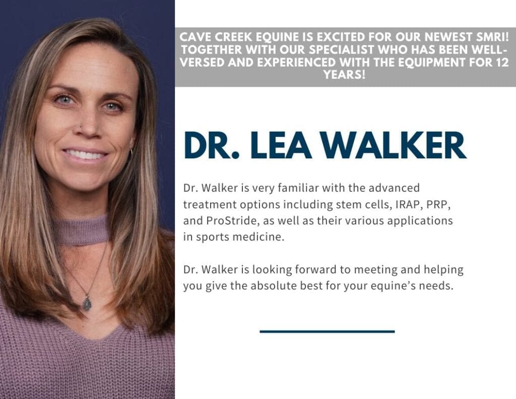
With 12 years of knowledge and experience in sports medicine, advanced imaging, and neurology, Dr. Walker is thrilled to help us attain our mission of becoming the premier sports medicine practice in the State of Arizona.
She is experienced in in-depth lameness examinations, advanced diagnostic imaging such as nuclear scintigraphy (bone scan), MRI, ultrasound, and digital radiography. Dr. Walker is also very familiar with all advanced treatment options including stem cells, IRAP, PRP and ProStride, etc. and their various applications in sports medicine. Her previous 10+ years of experience with using our newest equipment makes us even more exhilarated to offer this to our treasured clients.
Dr. Walker looks forward to meeting and helping you give the absolute best for your equine.


